• Vidhan Diagnostic Centre

Viability scan (Early Pregnancy Scan)

This is an ultrasound examination that is usually carried out vaginally between 6-10 weeks of pregnancy.

The aim of this scan is to determine the number of embryos present and whether the pregnancy is progressing normally inside the uterus.

This scan is useful for women who are experiencing pain or bleeding during the pregnancy and those who have had previous miscarriages or ectopic pregnancies.

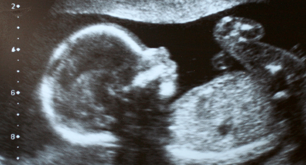
The figure on the left shows a normal pregnancy at 7 weeks of gestation. The figure on the right shows the empty sac of an anembryonic pregnancy at 7 weeks.

Nuchal scan (NT Scan)

This scan is carried out between 11 weeks to 13 weeks+6 days. The scan is usually performed transabdominally but in a few cases it may be necessary to do the examination transvaginally.

Anomaly scan

This is a detailed scan done between 20-24 weeks of pregnancy.

During the scan, we examine each part of the fetal body, determine the position of the placenta, assess the amount of amniotic fluid, and measure the fetal growth. Special attention is paid to the brain, face, spine, heart, stomach, bowel, kidneys and limbs.

If any abnormalities are detected, the significance of the findings will be discussed and the couple will be given the opportunity to have further counselling

Cardiac scan

During the nuchal scan (11-13 weeks), the anomaly scan (20-24 weeks) and wellbeing scan (30-34 weeks) we routinely examine the fetal heart and connecting blood vessels.

Women with a family history of congenital heart abnormalities, those with diabetes mellitus and those taking antiepileptic drugs.

Fetuses with suspected heart defect and those with increased nuchal translucency or certain non-cardiac abnormalities detected during the routine scans.

Wellbeing scan

This ultrasound scan is usually carried at about 32 weeks of pregnancy.

Some obstetricians advise that this scan is offered to all women. Others reserve such scans for those women who have had previous complications in pregnancy such as pre-eclampsia, growth restriction, diabetes, stillbirth, and for those women who develop a problem during the course of their current pregnancy.

Cervical scan

This is a transvaginal scan to measure the length of the cervix.

It is recommended for women at high risk for preterm birth, including multiple pregnancies, those with a previous preterm birth, abnormalities of the uterus or previous cervical surgery.

This examination is usually carried out at the time of the anomaly scan but in women with previous preterm birth it may be necessary to perform a series of scans starting from 16 weeks.